
-
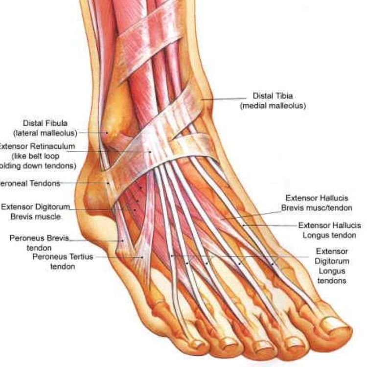
Foot & Ankle
Treatable Conditions
Achilles’ tendon injury
Chronic ankle laxity
Foot and ankle sprain
5th metatarsal fracture
Stress fractures
Metatarsalgia
Mortons neuroma
Over-Pronation
Planter Fasciitis
Spurs
Hallux rigidus
Metatarsal stress fracture
Heel fat pad pain syndrome
Entrapment of first branch of planter nerve & medial nerve
Peroneus brevis Tendon injury
Tarsal tunnel syndrome
Turf toe
Midtarsal sprain -
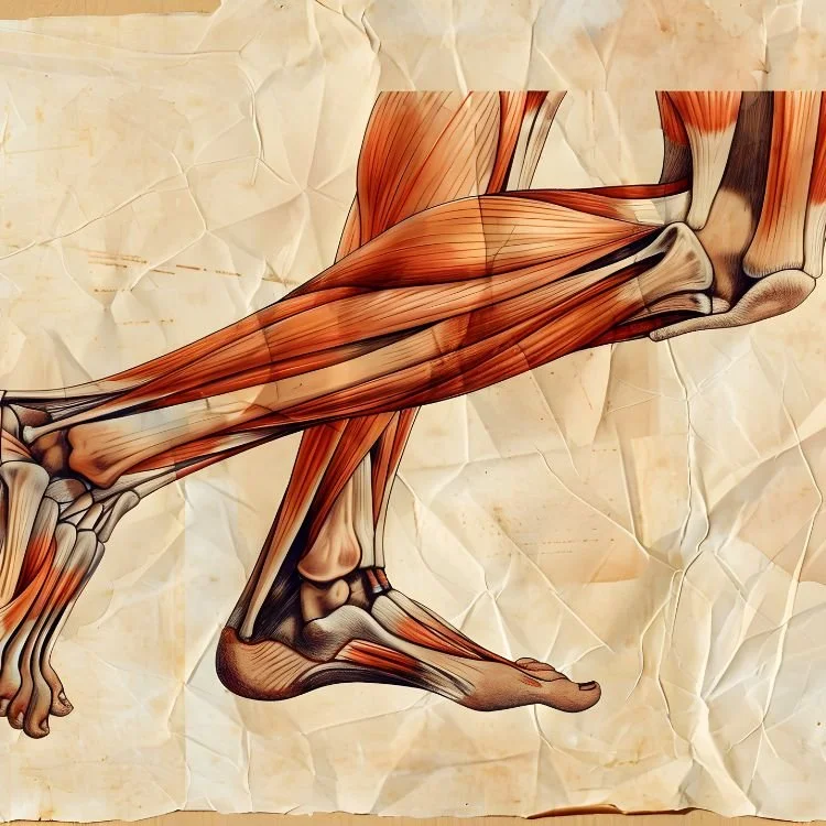
Leg
Treatable Conditions
Calf strain
Perineal tendon strain
Posterior tibial tendinitis
Shin splints
Anterior compartment syndrome
Posterior deep compartment syndrome
Medial tibial stress syndrome
Distal tibial syndrome
Plantar Longus injury
Common peroneal nerve injury
Lateral compartment syndrome -
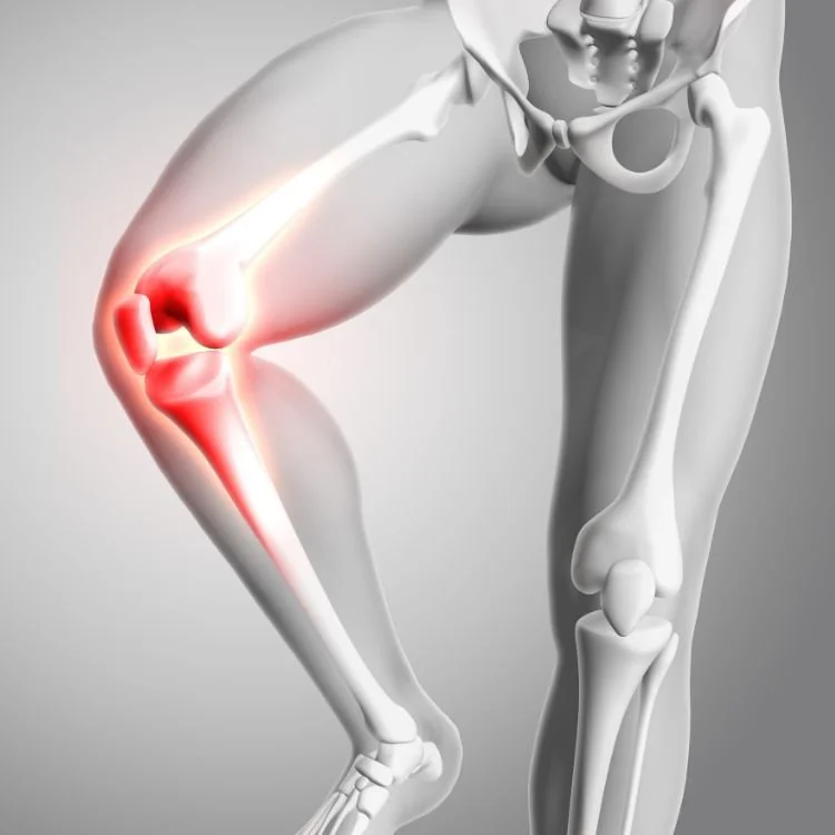
Knee
Treatable Conditions
Anterior cruciate ligament (ACL) injury
Baker Cyst
Ilioibial band syndrome
Lateral collateral ligament sprain
Medial collateral ligament sprain
Meniscal (cartilage) tear
Osgood-Slattery disease
Planter (kneecap) subluxation
Planter tendinitis (jumper's knee)
Patellofemoral pain syndrome (runner's knee)
Pes Anserine (knee) bursitis
Pelica syndrome
Sinding-Larsen-Johansson disease
Quadriceps tendon injury -
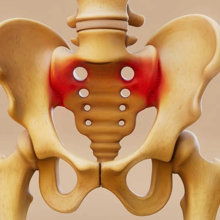
Hip, Thigh & Pelvis
Treatable Conditions
Gluteal strain
Gluteal tendinitis
Hamstring strain
Hip flexor strain
Hip pointer
Osteitis Pubis
Piriformis syndrome
Quadriceps contusion and strain
Snapping hip syndrome
Trochanteric bursitis
Iliopsoas overuse injury
Iliopsoas rupture
Hamstring syndrome
Traumatic myositis ossificans -
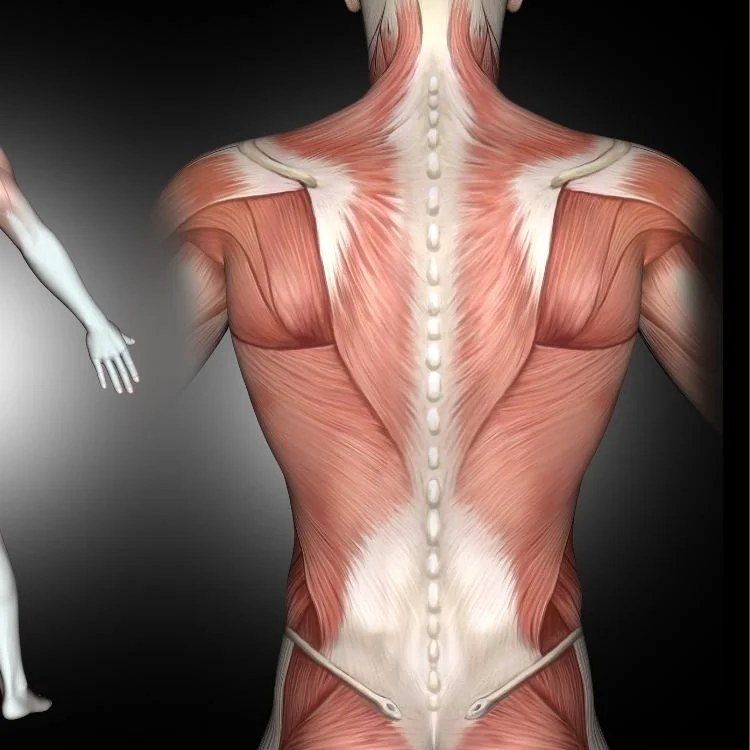
Back
Treatable Conditions
Herniated disk
Low back pain
Sacroiliac pain
Scoliosis
Spondylolysis
Spondylolisthesis
Tailbone injury
Upper back
Facet joint syndrome
Lumbago
Sciatica
Spinal sternosis
Weak back
Ankylosing spondylitis -
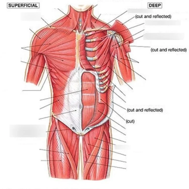
Chest & Adomin
Treatable Conditions
Abdominal muscle strain
Broken collarbone
Rib injury
Sternoclavicular joint separation -
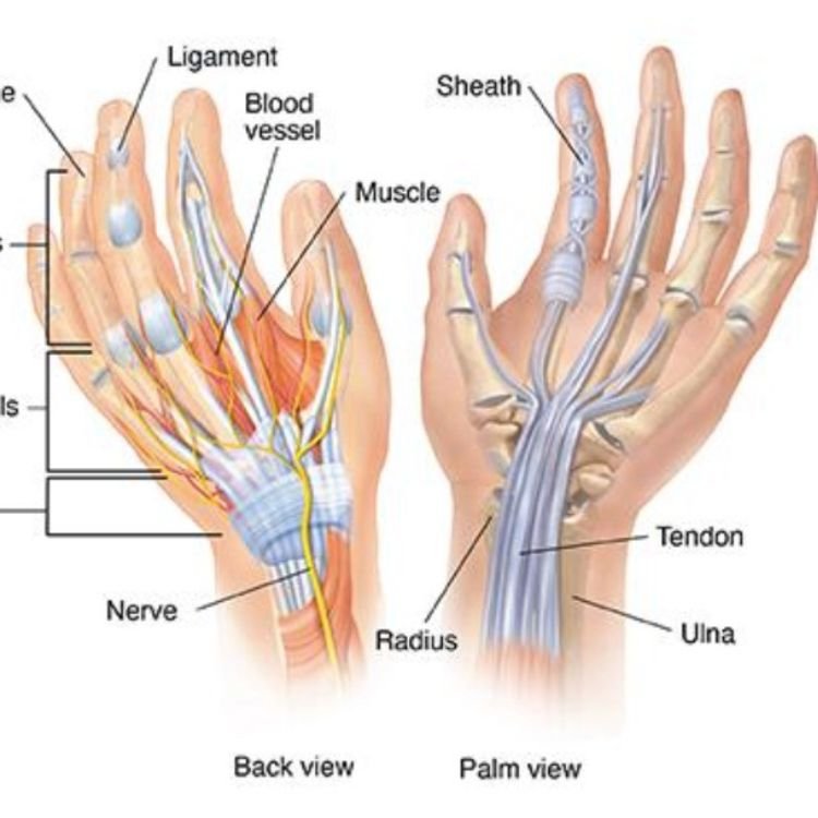
Wrist & Hand
Treatable Conditions
Carpel tunnel syndrome
Broken collarbone
Rib injury
Sternoclavicular joint separation
De Quervains tenosynovitis
Finger sprain
Thumb sprain
Triangular fibrocartilage complex (TFCC) injuries
Trigger finger
Ulnar collateral ligament sprain
Ulnar neuropathy
Wrist fracture
Wrist sprain
Wrist tendinitis -
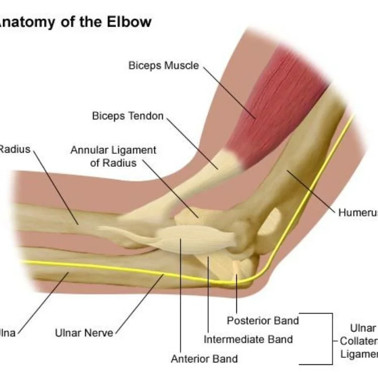
Elbow & Arm
Treatable Conditions
Elbow (Olecranon) bursitis
Lateral epicondylitis (tennis elbow)
Medial epicondylitis (golfers elbow)
Triceps tendinitis -
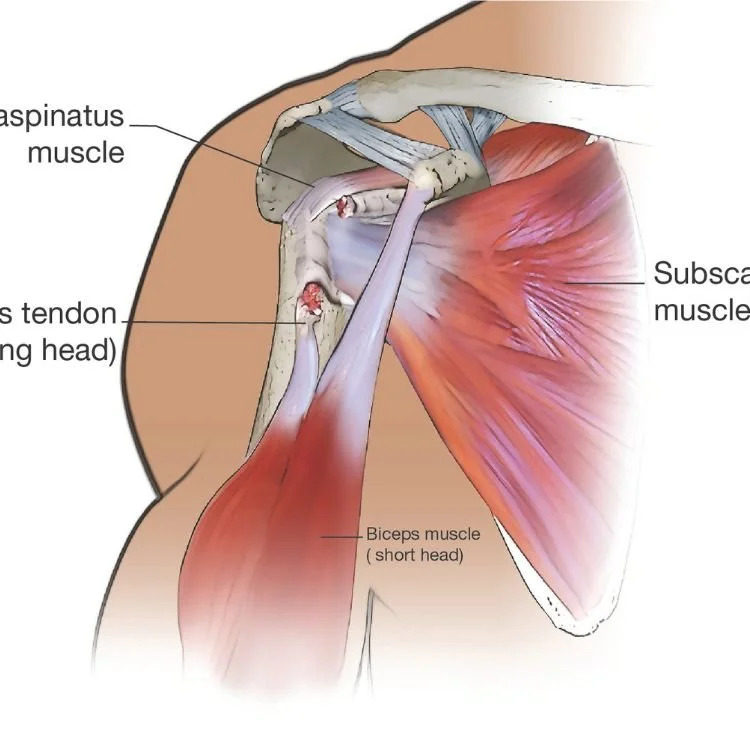
Shoulder
Treatable Conditions
Biceps Tendonitis
Frozen shoulder
Lateral tear of the shoulder
Rhomboid muscle strain and spasm
Rotator cuff injury
Shoulder bursitis -
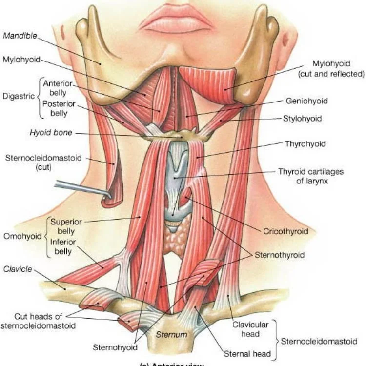
Neck
Treatable Conditions
Neck spasms
Neck strain
Lateral tear of the shoulder
Rhomboid muscle strain and spasm
Rotator cuff injury
Shoulder bursitis
Brachial plexus injury
Cervical brachialgia -
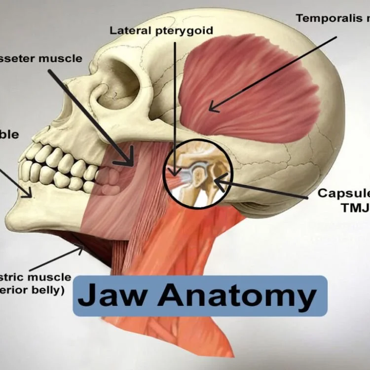
Jaw & Head
Treatable Conditions
Headaches
Greater Occipiral nerve injury
Migraine
Sinus pain
Occipital pain
Temporal mandibular joint dysfunction